


CT Dent’s latest case of the month on planning for dental implants includes; the main findings, findings from the radiographic impressions and images – displaying measurements and analysis.
It is also possible to view and manipulate this CBCT in CT Dent’s cloud viewer online.
CBCT scanner: Kavo OP 3D Vision V17
CBCT imaging protocol: 16cm height x 10cm width, 0.25mm voxel
Effective dose: 0.15 mSv
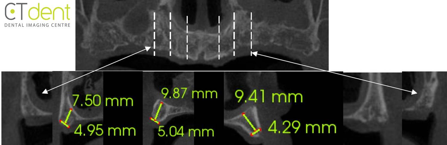
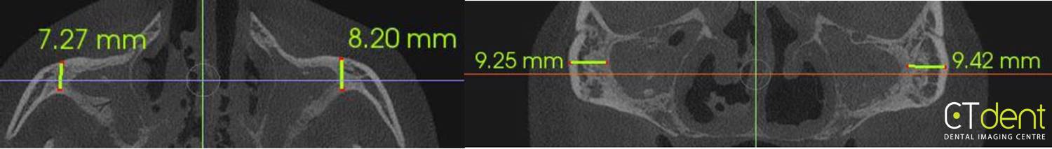
Clinical information: scan assessment prior to implants in mandible and zygoma.
Click here to view and manipulate this case of the month CBCT on CT Dent’s Cloud Viewer.
Main findings
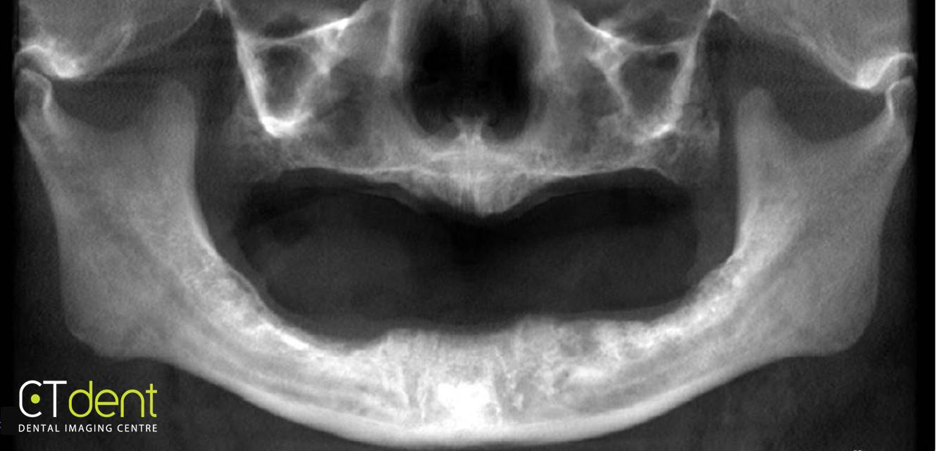
Maxilla: no abnormalities detected.
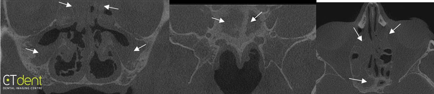
Sinuses: a generalised increase in the thickness and density of the tissues lining the right and left maxillary, ethmoid, and sphenoid sinuses were noted. The bone forming the maxillary and sphenoid sinuses is thickened and sclerotic/increased in density.
Nasal cavity: no abnormalities detected.
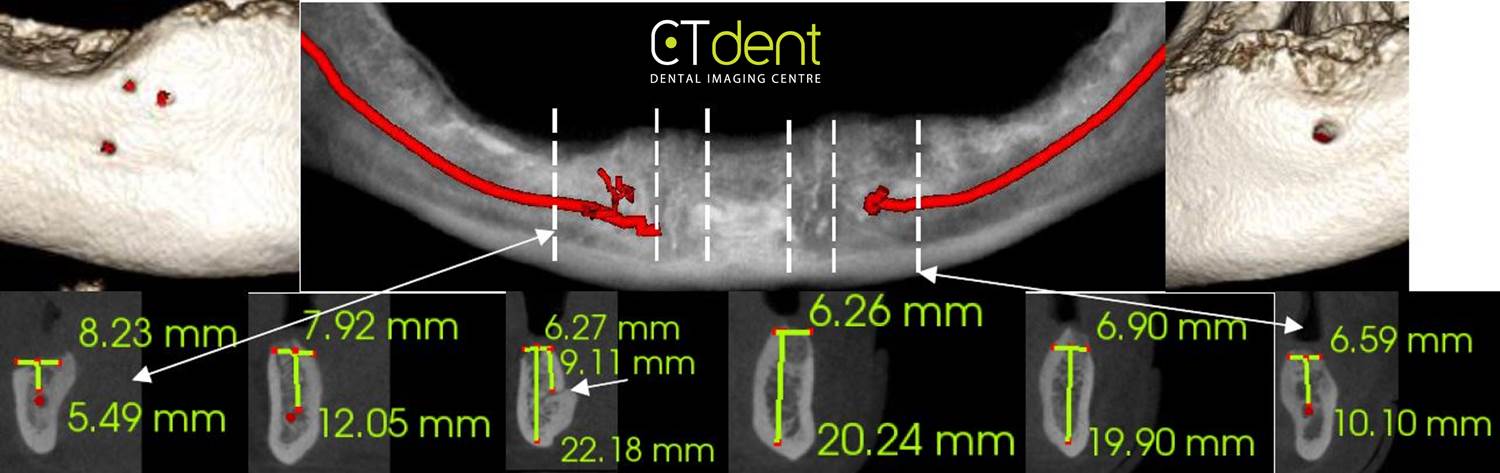
Mandible: no abnormalities detected.
Air space: mild enlargement of the pharyngeal tonsils was noted.
TMJs: no abnormalities detected; visible portions of the condyle exhibit a smooth continuously corticated outline and good symmetry.
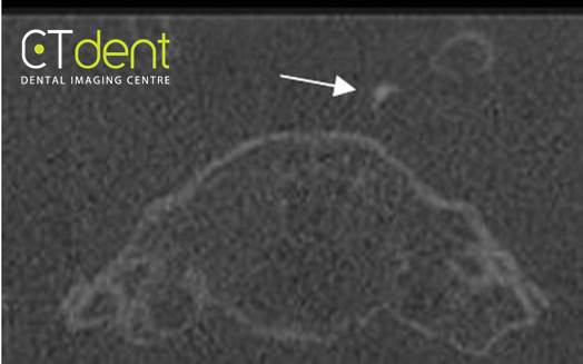
Other findings: curvilinear areas of increased density were noted lateral to the pituitary fossa in areas anatomically associated with the carotid arteries; these areas appear to be consistent with calcification of the carotid artery.
Dental findings: no abnormalities detected.
Radiographic impression
Sinuses: the radiographic findings are consistent with a generalised, long-standing chronic mucositis/sinusitis of the right and left maxillary sinuses. The thickened, sclerotic walls of the maxillary and sphenoid sinuses are an indication of long-standing chronic inflammatory changes. Correlation of the radiographic observation with the patient’s clinical history and symptoms if any is suggested. Physician referral for more thorough evaluation is suggested.
Airspace: clinical evaluation of the soft tissues of the oropharynx is suggested.
Other findings: vascular calcifications may be an indication of a potential increased risk of cardiovascular disease and stroke. Correlation of the radiographic observation with the patient’s clinical history of high blood pressure, elevated cholesterol, smoking and stress is suggested. Physician referral for more thorough evaluation is suggested if merited by clinical findings.
The following are selected images from the volume illustrating major findings.
The importance of CBCT in implant dentistry
Since its introduction for dental and maxillofacial imaging, cone beam CT has played an important role alongside clinical evaluation with the planning and follow-up of dental implant treatment.
Limited or full-volume fields of view can be used to determine bone volume and quality and the shape of the alveolar ridge.
For the mandible it can be used to check the position of the inferior dental canal, mental foramen and anterior looping and salivary gland depressions.
In the maxilla, assessment can be made of the nasopalatine canal, nasal fossa and the maxillary sinuses.
Although sometimes limited by restoration noise and artifact, lower dose CBCT post implant placement can be an invaluable tool for checking implant position, any close relationship with important anatomical structures and peri-implantitis.
CT Dent provides a dental imaging service in seven UK centres, where all scans are taken by qualified radiographers using the latest CBCT scanners to ensure high quality imaging and patient satisfaction.
For more information on our services please visit www.ct-dent.co.uk or call 020 7487 5717.
Let’s block ads! (Why?)
