 Chang IC Teoh demonstrates the clinical effectiveness of regenerating vertical bone volume.
Dental implant therapy has been established as a predictable treatment option of restoring partially edentulous mouths (Pjetursson et al, 2012).
One of the prerequisite conditions for successful treatment is having sufficient bone volume for optimal three dimensional implant placement (Resnik and Misch, 2017).
Many clinical situations present with inadequate bone volume. They require bone augmentation of varying degrees, with or without simultaneous implant placement.
For many clinicians, ridge augmentation of medium to large vertical defects is often considered to be challenging. Many techniques (Urban et al, 2014; Jensen and Terheyden, 2009; Simion and Trisciuoglio, 2016; Jenson et al, 2002; Khoury and Hanser, 2015; Sahjheb et al, 2017) have been described for the augmentation of vertical ridge defects.
GBR
These techniques include GBR (guided bone regeneration) using a combination of membrane barriers and different grafting materials; autogenous bone block grafts; autogenous particulate grafts; 3D split bone block; distraction osteogenesis; 3D CAD/CAM titanium scaffolds with different types of biomaterials; or a combination of these.
The dimensional changes of bone following tooth loss have been well documented (Araujo and Lindhe, 2005; Chappuis et al, 2013; Chappuis et la, 2013). Final ridge morphology is often the result of a combination of predisposing factors influencing the area affected.
Bone destruction caused by advanced periodontitis, multiple tooth loss, peri-implantitis, trauma, long-term denture wearing, unfavourable loading, or a combination of these factors may result in advanced bone loss and severe ridge defects in either horizontal, vertical or a combination of dimensions (Atwood, 2001; Tonetti et al, 2018; BDIZ/EDI, 2013).
There are a number of classifications of bony defect described in the literature. The Cologne classification of alveolar ridge defects (CCARD) (BDIZ/EDI, 2013) offers a more comprehensive description of the types of ridge defect presented to the clinicians and their management.
VRA vs. HRA
In general, vertical ridge augmentation (VRA) is more demanding in soft tissue management, and the need to stabilise the augmentation material, than horizontal ridge augmentation (HRA).
The inclusion of autogenous bone materials in vertical ridge augmentation (VRA) is often recommended and improves the outcome. However the complication rates (Fontana et al, 2011; Fontana et al, 2008; Tinti and Parma-Benfenati, 1998) associated with VRA are considerably higher compare with horizontal ridge augmentation (HRA).
VRA is challenging primarily due to the difficulty of stabilising the bone graft material without the support of the bony wall and angiogenesis having to reach a distance from the native bony bed (Urban et al, 2019; Urban, 2017).
In addition, an absolute tension-free soft tissue advancement is essential to achieve primary closure and prevent wound dehiscence during the entire period of healing (Urban et al, 2019; Urban, 2017).
The author would like to present vertical ridge augmentation (VRA) cases in a series of articles using different VRA techniques.
Case study

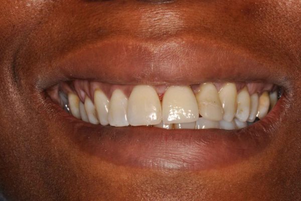
A 30 year-old Afro-Caribbean lady with good health presented with a mobile UL1 in 2017. She worked as a schoolteacher and her past dental history revealed that the UL1 and UL2 suffered from a traumatic injury when she was a teenager.
The UL1 was avulsed and successfully reimplanted at the time of injury by her dentist. The dentist restored the fractured UL2 with a partial veneer.
Both teeth were uneventful after the treatment, but the UL1 had started to become increasingly mobile over the last few years.
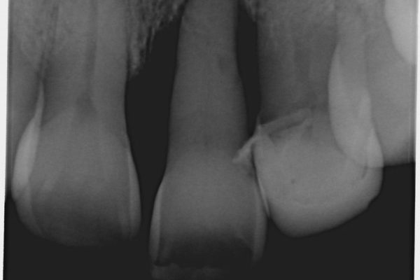
Periapical radiograph showed that the UL1 had advanced bone loss with poor prognosis (Figure 1). The patient wanted a fixed implant treatment option to replace the failing UL1.
After careful discussion with the patient, a treatment plan was formulated. A staged approach to rehabilitating the aesthetically demanding area was chosen.
Stage 1:
Removal of the UL1 and temporary replacement with metal acrylic adhesive bridge. Reassessment after three months of healing.
Stage 2:
VRA with GBR technique using titanium reinforced cytoplast d-PTFE membrane and Emdogain (enamel matrix derivatives) treatment.
Stage 3:
Implant placement eight months after VRA. Soft tissue augmentation, with provisional crown and final crown on implant.
The clinical presentation after extraction of the UL1 was as follows:
- Medium to high lip line
- Thin biotype
- Medium vertical bone defect
- Papilla loss mesial UL2
- Reduced interproximal bone peak mesial to UL2
- Class I incisal relationship
- UL2 is restored with a partial porcelain veneer
The ridge defect was diagnosed with a Cologne classification of V.2.i – a vertical defect of 4-8mm, inside the ridge contour.
Extraction
The failing UL1 was extracted and an adhesive temporary Maryland bridge was cemented to neighbouring teeth. The area was allowed to heal for three months: this would allow the soft tissue time to mature and revascularisation of the area. The healing was uneventful.
Reassessment of the alveolar ridge defect revealed both of the palatal bone plates were missing (Figures 2-5) with a vertical bone defect of around 5mm in the deepest. In general, vertical defects of over 4mm suggest a staged approach should be more indicated.
Guided bone augmentation was carried out as described by Urban (Urban et al, 2014 Urban, 2017).
A crestal incision was made and the releasing incision was made at least one tooth behind the defect. A full thickness three-sided flap was raised to expose the full extent of the bony defect. The bony defect was mainly in the vertical dimension and moderate in size (around 5mm): both buccal and palatal bone was missing (Figures 6 and 7).
Bony peaks were clearly present on the mesial part of the UR1 and UL2, which determined the vertical limit of bone regeneration. Adequate bone width was available to support grafting material.
Augmentation
The granulation tissue was removed: it is important not to leave any soft tissue remnants in the area to be augmented. At the same visit, the exposed root surface of the UL2 was cleaned, curetted and enamel matrix derivatives applied in order to promote tissue regeneration on the root surface.
Autogenous bone chips were harvested from the anterior mandible with a 6mm diameter trephine (Figure 8). Autogenous bone chips provide material of excellent osteogenic properties for bone regeneration.
The harvested autogenous bone was mixed with xenograft (Bio-Oss particles), at a ratio of around 50:50, to increase the bulk volume of the grafting material.
A non-resorbable titanium reinforced d-PTFE (cytoplast) membrane was used to protect the grafting material and at the same time offered increased mechanical stability for rebuilding the ridge defect (Figure 9).
Two titanium fixation pins were placed to stabilise the membrane (Figure 10).
A resorbable collagen membrane (Bio-Gide) was placed over the titanium reinforced d-PTFE membrane and the grafting material that was not covered by the d-PTFE membrane (Figure 11).
The flap was rendered tension-free by dividing the periosteum at the base of the buccal flap with one single incision and the underlying connective tissue was further stretched with a blunt instrument. The flap was then closed with d-PTFE sutures. A postoperative radiograph was taken to provide a baseline record (Figure 12).
Implant placement
The area was allowed to heal for eight months, which were uneventful. Throughout that period, the patient functioned well with the adhesive temporary bridge.
The ridge defect and the soft tissue attachment mesial to the UL2 were markedly improved, although there was still a small gap present between the intaglio surface of the temporary bridge and the tissue surface (Figure 13). A connective tissue graft at implant surgery would correct this defect.
A full thickness flap was raised and the non-resorbable membrane was removed (Figures 14 and 15). The newly regenerated bone was impressive, with good volume, and a dental implant (Straumann BLT RC Roxolid implant) was placed in the correct three dimensional position with good primary stability (Figures 16 and 17).
A piece of connective tissue graft was harvested from the palate to increase the tissue volume in the crestal region (Figure 18).
Primary closure was achieved, and healing was uneventful. The increase in soft tissue volume eliminated the gap existed between the intaglio surface of temporary bridge and the tissue surface of the ridge (Figure 19).
Prosthetic restoration
The implant was exposed two months after insertion with a H-incision technique and minimal soft tissue manipulation. The papillary area was left undisturbed (Figure 20).
An impression was then taken after implant exposure, and a temporary crown was fabricated with the correct emergence profile to encourage soft tissue ingrowth and moulding around it (Figure 21).
The soft tissue was supported by a stable hard tissue foundation of sufficient volume. That would encourage proper soft tissue development around the implant including the lost papilla tissue mesial to the UL2 (Figures 22 and 23).
A final impression was taken once the soft tissue moulding was matured. A screw-retained all-ceramic crown was then delivered with a good aesthetic result (Figures 25-26). The patient was very pleased with the final result.
Conclusion
This article presented the treatment of a highly aesthetically demanding case with a vertical bone defect.
A staged approach of tissue regeneration was selected. Firstly, hard tissue regeneration was applied using the principle of guided bone regeneration (autogenous bone chips and xenograft protected by titanium reinforced d-PTFE membrane), and Emdogain was used to regain lost attachment next to a natural tooth. Both stages were meticulously executed.
It is important to use a mechanically stable membrane such as titanium-reinforced d-PTFE membrane to provide a closed and undisturbed environment in which the bone graft can mature. Sufficient time must also be allowed for the hard tissue to be regenerated.
At implant placement, soft tissue volume regeneration was carried out using connective tissue graft to increase the bulk volume of soft tissue in the crestal region.
It is essential to regenerate sufficient volume of hard tissue both in vertical and horizontal dimension – not only to house and surround the implant inserted (2-4mm of buccal bone) (Spray et al, 2000; Grunder et al, 2005), but also to provide a good foundation for the soft tissue to be adequately supported.
It is now known that adequate vertical soft tissue volume is important for long term stability of crestal bone around dental implants (Linkevicius, 2019).
Last but not least is the design of the emergence profile of the provisional and final restorations: they are the key to developing and maintaining the correct soft tissue profile with sufficient volume and thereby achieving a predictable good aesthetic final outcome.

For references, contact: [email protected]
This article first appeared in Implant Dentistry Today magazine. You can read the latest issue here.
Let’s block ads! (Why?)


